
Science Community
In every science activity, I benefit both as a presenter by further understanding my own science from the perspectives of my audience and as a listener by learning new science from my intelligent colleagues.
I enjoy communicating my science using different languages when facing different audiences and building platforms to facilitate better science communication.
Building science communication platforms
Imaging science student seminar initiator and co-chair
 WashU has PhDs from many departments and they are working on various imaging-related research, e.g., MRI, CT, OCT, fluorescence imaging, machine learning on imaging …
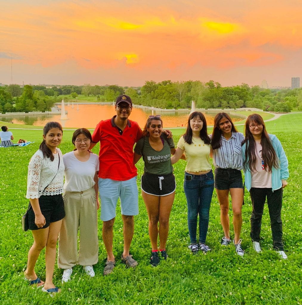
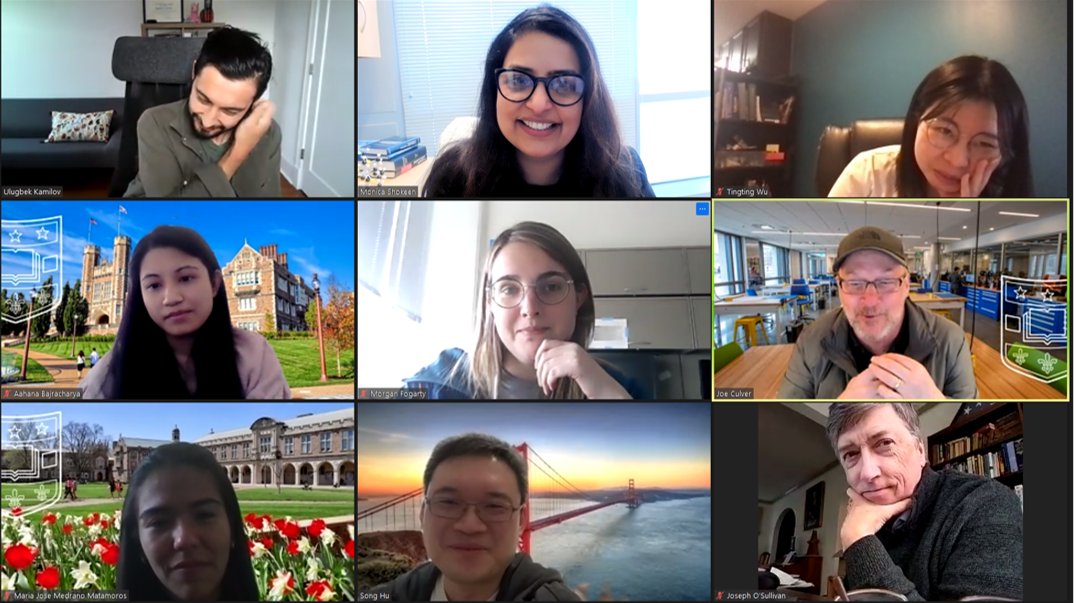
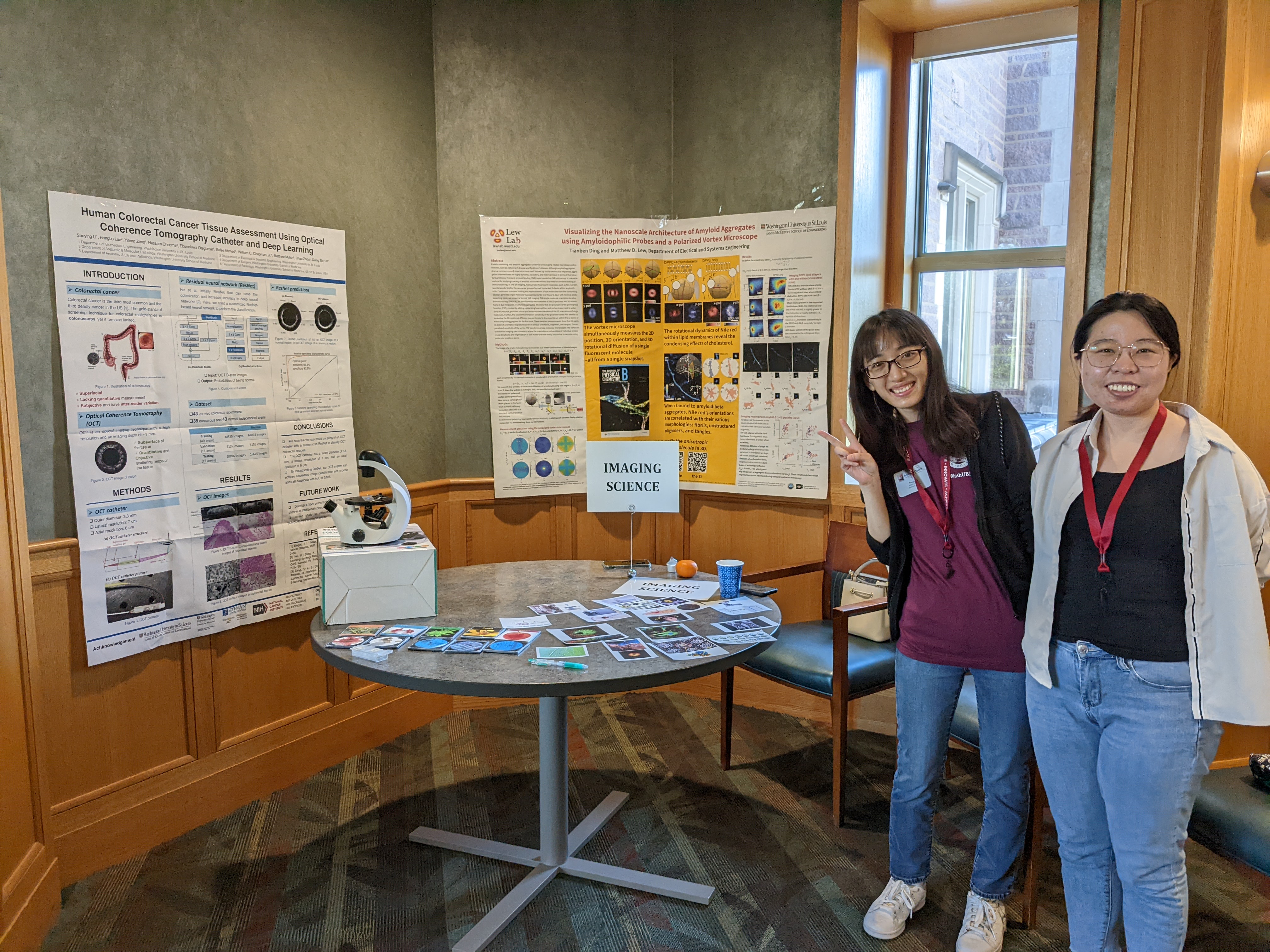
Together with Aahana Bajracharya, Maria Medrano, and Chunhui Yang, we initiated monthly imaging science student seminars starting in the fall of 2019. We invited Ph.D. students from different programs to share their awesome imaging research with our audiences. Till the end of April 2022, we already hold around 20 seminars.
During the pandemic time, we decided to build our ‘imaging science library’ where we shared our presenters’ presentations after their seminars. We hope this library will help new students find the labs they are interested in, and help existing students know more about the imaging science happening at WashU.
Imaging science pathway retreat committee
 As a committee member, I helped to organize the annually imaging science pathway retreat in 2020. Learning and participating in the organization and planning process of a big conference is beneficial.
Science outreach
St. Louis science center outreach
Lew lab had a science table at the St. Louis Science Center for demonstrating Lewlab’s research to more than ~200 visitors. We demonstrated 1) how we leverage fluorescence to see things in the dark, and 2) how we achieve high resolution using single-molecule imaging through a playing game.
Science coach program in WashU
 I am a science coach in the event organized by Association of Graduate Engineering Students (AGES) at WashU.
We meet high school students and facilitate them to brainstorm research projects. I am flattered that one student was inspired by our imaging demo and come up with an idea of how the color of stars is related to their distance to the center of the milky way galaxy.
Invited research speaker for math camp in WashU
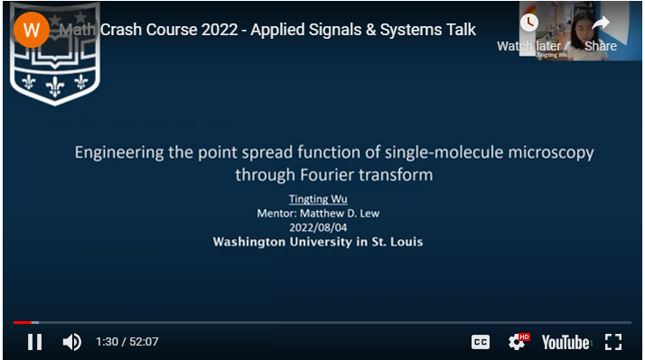 I gave a talk titled “Engineering the point spread function of single-molecule microscopy through Fourier transform” on Math Camp in WashU for the incoming Ph.D. students in the imaging science program.
This is my first time giving a lecture about Fourier optics, and I was thrilled to share this beautiful physics and mathematics with the new students.
I gave a talk titled “Engineering the point spread function of single-molecule microscopy through Fourier transform” on Math Camp in WashU for the incoming Ph.D. students in the imaging science program.
This is my first time giving a lecture about Fourier optics, and I was thrilled to share this beautiful physics and mathematics with the new students.
Tingting’s research on News
Machine learning generates pictures of proteins in 5D
Lew lab sheds new light on cell membranes
New microscopy method provides unprecedented look at amyloid protein structure
Science artworks
Journal Arts
Where do these Journal Arts come from?
For some of my journal publications, I tried to create art images to submit for journal covers. Whenever I do those journal art designs, I find myself too passionate and even stay super late to create those images. A kind of passion never occurs when I am writing papers or working on projects :). Unfortunately, none of them have been selected as cover images for the journal, so I decided to put them here.
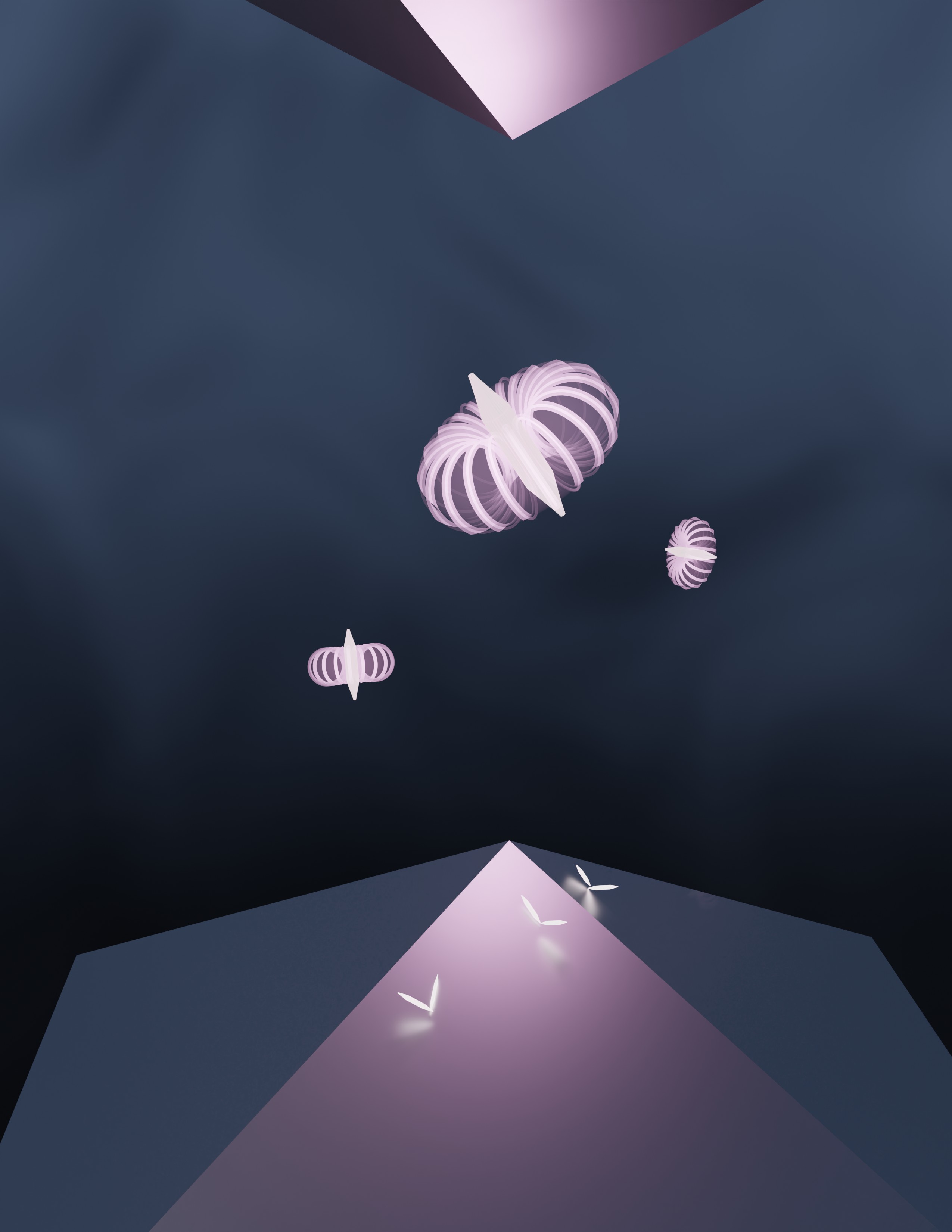 A novel imaging system design using two pyramid mirrors, termed raMVR. Dipole-like fluorescent dyes (structure at the middle) are imaged using this optical system. [created using Blender]
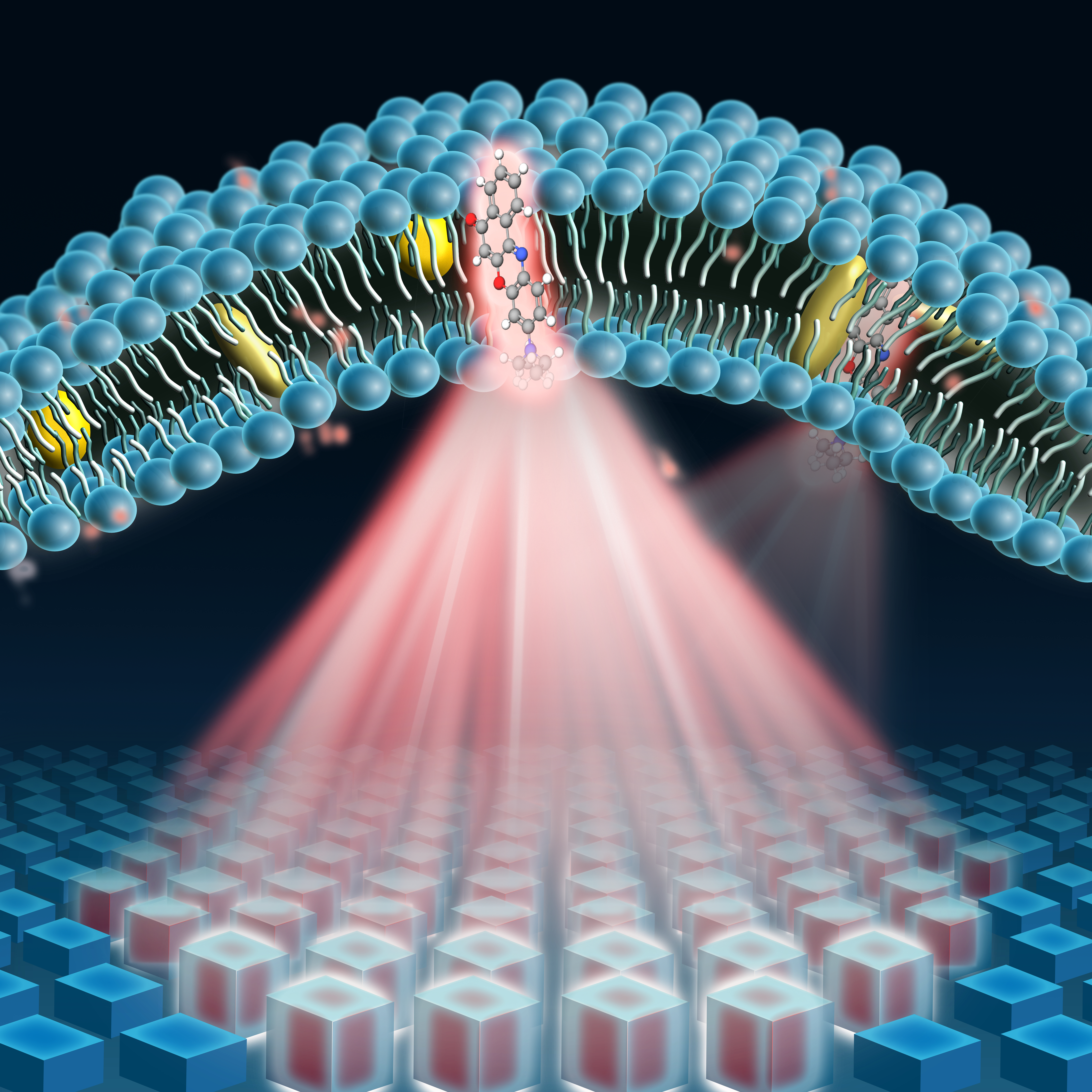 Nile red, a fluorescent emitter, transiently binds to the lipid bilayers at a certain orientation and emit fluorescence light. Our pixel-wise optimized phase mask is placed at the back focal plane of the microscope to modulate the phase of fluorescence light. After modulation, the shape of point spread function enables us to measure the 3D orientation and 3D position of Nile red simultaneously. [created using Affinity designer]
WashU engineering school calendar
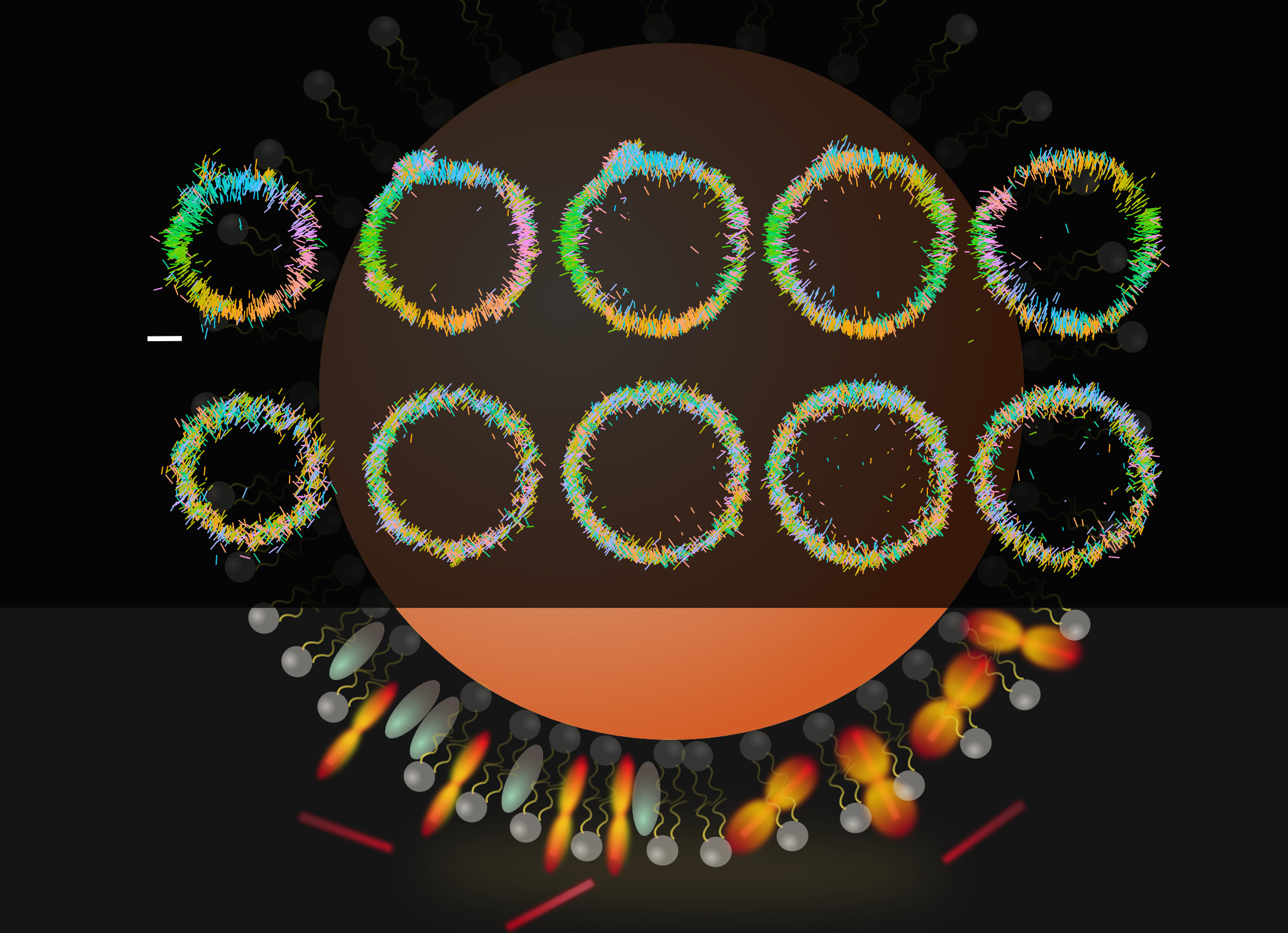 The pixOL microscope enables super-resolution imaging of the 3D locations and 3D orientations of fluorescent molecules. The 6-dimensional images from pixOL depict Nile red molecules (NR, red bars) as they transiently bind to a spherical lipid bilayer (SLB, gray structures around the sphere). The top two rows show the orientations of NR (each line represents the long axis of the molecule) at different z-slices through the sphere (left to right). With cholesterol (aqua ellipses) present in the SLB, we observe that NR becomes more ordered and perpendicular to the sphere (top row); without cholesterol present to organize the membrane, NR exhibits various orientations (second row). Scale bar: 400 nm. [created using Affinity designer]